The important structures of the elbow can be divided into several categories. These include
- bones and joints
- ligaments and tendons
- muscles
- nerves
- blood vessels
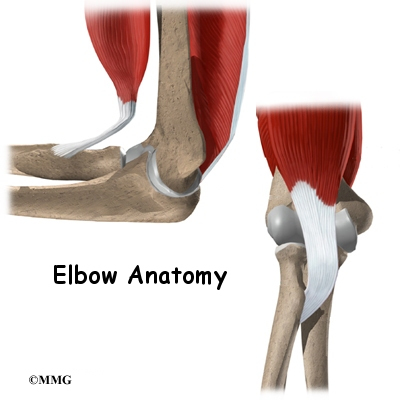
Bones and Joints
The bones of the elbow are the humerus (the upper arm bone), the ulna (the larger bone of the forearm, on the opposite side of the thumb), and the radius (the smaller bone of the forearm on the same side as the thumb). The elbow itself is essentially a hinge joint, meaning it bends and straightens like a hinge. But there is a second joint where the end of the radius (the radial head) meets the humerus. This joint is complicated because the radius has to rotate so that you can turn your hand palm up and palm down. At the same time, it has to slide against the end of the humerus as the elbow bends and straightens. The joint is even more complex because the radius has to slide against the ulna as it rotates the wrist as well. As a result, the end of the radius at the elbow is shaped like a smooth knob with a cup at the end to fit on the end of the humerus. The edges are also smooth where it glides against the ulna.
Articular cartilage is the material that covers the ends of the bones of any joint. Articular cartilage can be up to one-quarter of an inch thick in the large, weight-bearing joints. It is a bit thinner in joints such as the elbow, which don't support weight. Articular cartilage is white, shiny, and has a rubbery consistency. It is slippery, which allows the joint surfaces to slide against one another without causing any damage.
The function of articular cartilage is to absorb shock and provide an extremely smooth surface to make motion easier. We have articular cartilage essentially everywhere that two bony surfaces move against one another, or articulate. In the elbow, articular cartilage covers the end of the humerus, the end of the radius, and the end of the ulna.
Ligaments and Tendons
There are several important ligaments in the elbow. Ligaments are soft tissue structures that connect bones to bones. The ligaments around a joint usually combine together to form a joint capsule. A joint capsule is a watertight sac that surrounds a joint and contains lubricating fluid called synovial fluid.
In the elbow, two of the most important ligaments are the medial collateral ligament and the lateral collateral ligament. The medial collateral is on the inside edge of the elbow, and the lateral collateral is on the outside edge. Together these two ligaments connect the humerus to the ulna and keep it tightly in place as it slides through the groove at the end of the humerus. These ligaments are the main source of stability for the elbow. They can be torn when there is an injury or dislocation to the elbow. If they do not heal correctly the elbow can be too loose, or unstable.
There is also an important ligament called the annular ligament that wraps around the radial head and holds it tight against the ulna. The word annular means ring shaped, and the annular ligament forms a ring around the radial head as it holds it in place. This ligament can be torn when the entire elbow or just the radial head is dislocated.
There are several important tendons around the elbow. The biceps tendon attaches the large biceps muscle on the front of the arm to the radius. It allows the elbow to bend with force. You can feel this tendon crossing the front crease of the elbow when you tighten the biceps muscle.
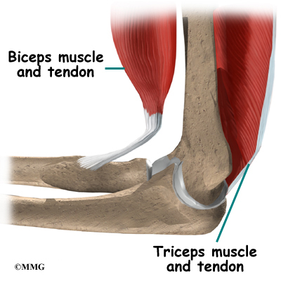
The triceps tendon connects the large triceps muscle on the back of the arm with the ulna. It allows the elbow to straighten with force, such as when you perform a push-up.
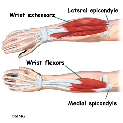
The muscles of the forearm cross the elbow and attach to the humerus. The outside, or lateral, bump just above the elbow is called the lateral epicondyle. Most of the muscles that straighten the fingers and wrist all come together in one tendon to attach in this area. The inside, or medial, bump just above the elbow is called the medial epicondyle. Most of the muscles that bend the fingers and wrist all come together in one tendon to attach in this area. These two tendons are important to understand because they are a common location of tendonitis.
Muscles
The main muscles that are important at the elbow have been mentioned above in the discussion about tendons. They are the biceps, the triceps, the wrist extensors (attaching to the lateral epicondyle) and the wrist flexors (attaching to the medial epicondyle).
 Nerves
All of the nerves that travel down the arm pass across the elbow. Three main nerves begin together at the shoulder: the radial nerve, the ulnar nerve, and the median nerve. These nerves carry signals from the brain to the muscles that move the arm. The nerves also carry signals back to the brain about sensations such as touch, pain, and temperature.
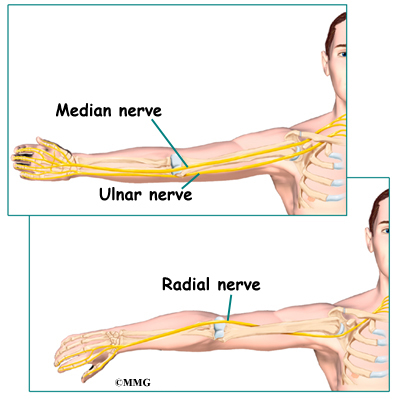
Some of the more common problems around the elbow are problems of the nerves. Each nerve travels through its own tunnel as it crosses the elbow. Because the elbow must bend a great deal, the nerves must bend as well. Constant bending and straightening can lead to irritation or pressure on the nerves within their tunnels and cause problems such as pain, numbness, and weakness in the arm and hand.
Blood Vessels
Traveling along with the nerves are the large vessels that supply the arm with blood. The largest artery is the brachial artery that travels across the front crease of the elbow. If you place your hand in the bend of your elbow, you may be able to feel the pulsing of this large artery. The brachial artery splits into two branches just below the elbow: the ulnar artery and the radial artery that continue into the hand. Damage to the brachial artery can be very serious because it is the only blood supply to the hand.